Ultrasound utilises sound waves to produce images of the body. A transducer is placed on the skin, which generates high frequency sound. The sound penetrates the body and is returned as echoes at the interface of anatomic structures or areas of pathology. The echoes are received by the transducer and are combined to form an image. Ultrasound is particularly useful for muscle and tendon injuries, particularly of the shoulder.
Portable Office Ultrasound, which is performed at the time of first consultation, offers the following advantages to patients:
Ultrasound can reveal many different diseases of the shoulder:

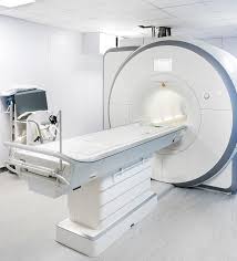
MRI Scans (short for Magnetic Resonance Imaging) is a powerful imaging technique which exploits the magnetic properties of hydrogen atoms within the body. This imaging technique uses magnetic fields and radio waves and does not employ ionizing radiation which normal x-rays use.
MR scanning does not use x-rays and there are no known harmful effects. MR scans show soft tissues such as muscles, ligaments, tendons and cartilage extremely well. CT scans and x-rays are better for viewing bones. Since most sports injuries involve the muscles, tendons and ligaments MR scanning is extremely useful.

WhatsApp us